

Cytotoxic lesion of the corpus callosum as presenting neuroradiological manifestation of COVID-2019 infection. Forestier G, de Beaurepaire I, Bornet G, Boulouis G. J Neurol. 2020 Aug 18;. doi: 10.1007/s00415-020-10166-1. [Epub ahead of print] PubMed PMID: 32809155; PubMed Central PMCID: PMC7433278.
Since December 2019, severe acute respiratory syndrome (SARS) cases related to the coronavirus disease-2019 (COVID-19) emerged from Wuhan, Hubei Province, China [1–3] and spread all around the world contaminating more than 17 million people (> 174,000 cases in France) [4]. Severe acute respiratory syndrome coronavirus-2 (SARS-CoV-2) leads to a wide spectrum of mild disease most of the time with respiratory system being the most commonly affected organ. Still, coronaviruses have an established neuroinvasive propensity [5, 6], and MRI findings associated with acute neurological manifestations in COVID-19 patients have been recently reported [7, 8] to explain to some extent the neurological symptoms of patients infected with the SARS-CoV-2.
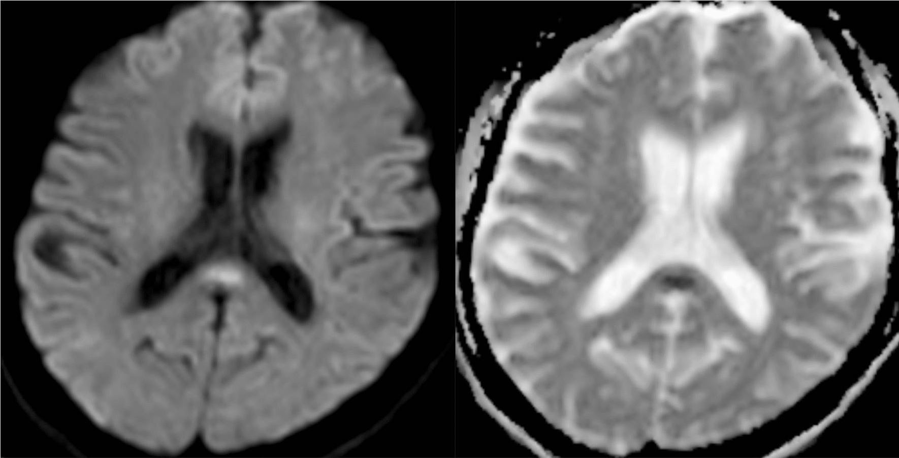
CLOCCs (previously termed Mild Encephalitis/Encephalopathy with Reversible Splenial lesion: MERS) is a rare infection-associated encephalopathy, involving cell-cytokine interactions [9], commonly described in the context of virus infections, metabolic disturbances, or antiepileptic drug. This syndrome presenting with great clinical heterogeneity [10], and complete regression of symptoms [11]. We hereby report the first case of cytotoxic lesion of the corpus callosum (CLOCCs) as presenting neuroradiological manifestation of COVID-2019 infection confirmed by chest computed tomography and nasopharyngeal swab sample test.
This part of the website could either be in French or English, depending on the sources of the actualities.