

Team leader : Catherine Oppenheim
Team member : Wagih BEN HASSEN | Grégoire Boulouis | Sylvain Charron | Clément Debacker | Myriam Edjali-Goujon | Anton François | Laurence Legrand | Julia Mathan | Jean-François Meder | Charles Mellerio | Olivier Nagarra | Sixtine Omont | Johan Pallud | Marion Plaze | Guillaume Raybaud | Alexandre Roux | Denis Trystram | Pascale Varlet | Marc Zanello
Key contributors : Elsa Angelini | Isabelle Bloch | Arnaud Cachia | Philippe Ciuciu | Joan-Alexis Glaunes | Pietro Gori | Nicolas Lomenie | Bertrand Thirion
 |
Together, the three axes cover a spectrum of pathological conditions, each with different time scales: acute onset (stroke), rapid or slow progression (Brain tumors), or lifelong evolution (mental disorders). We use state-of-the-art MRI acquisition and image processing techniques as well as advanced data analytics approaches including machine learning and multiscale imaging to achieve these goals. The team members, who share interest in MR biomarkers, have multidisciplinary skills: psychiatrists, neuropathologists, neurosurgeons, neuroradiologists, cognitive neuroscientists, research engineers with skills in physics of MR, micro-structural and fMRI analysis. A synergy between the three clinical axes fosters, in an interdisciplinary context, the sharing of methods for developing imaging biomarkers and theoretical approaches across application domains.
|

Main application axes
Mental disorders Imaging
Cachia A, Cury C, Brunelin J, Plaze M, Delmaire C, Oppenheim C, Medjkane F, Thomas P, Jardri R. Deviations in early hippocampus development contribute to visual hallucinations in schizophrenia. Transl Psychiatry. 2020;10:102. doi: 10.1038/s41398-020-0779-9.
Moyal M, Haroche A, Attali D, Dadi G, Raoelison M, Le Berre A, Iftimovici A, Chaumette B, Leroy S, Charron S, Debacker C, Oppenheim C, Cachia A, Plaze M. 2023. Orbitofrontal sulcal patterns in catatonia. European psychiatry 1–25. Advance online publication. https://doi.org/10.1192/j.eurpsy.2023.2461.
Neurovascular Imaging.
Benzakoun J, Deslys MA, Legrand L, Hmeydia G, Turc G, Hassen WB, Charron S, Debacker C, Naggara O, Baron JC, Thirion B, Oppenheim C. Synthetic FLAIR as a Substitute for FLAIR Sequence in Acute Ischemic Stroke. Radiology. 2022;303:153-159. doi: 10.1148/radiol.211394. Link Article:
Kerleroux B, Benzakoun J, Janot K, Dargazanli C, Eraya DD, Ben Hassen W, Zhu F, Gory B, Hak JF, Perot C, Detraz L, Bourcier R, Aymeric R, Forestier G, Marnat G, Gariel F, Mordasini P, Seners P, Turc G, Kaesmacher J, Oppenheim C, Naggara O, Boulouis G. Relevance of Brain Regions' Eloquence Assessment in Patients With a Large Ischemic Core Treated With Mechanical Thrombectomy. Neurology. 2021;97:e1975-e1985. doi: 10.1212/WNL.0000000000012863.
Neuro-Oncology.
Roux A, Roca P, Edjlali M, Sato K, Zanello M, Dezamis E, Gori P, Lion S, Fleury A, Dhermain F, Meder JF, Chrétien F, Lechapt E, Varlet P, Oppenheim C, Pallud J. MRI Atlas of IDH Wild-Type Supratentorial Glioblastoma: Probabilistic Maps of Phenotype, Management, and Outcomes. Radiology. 2019;293:633-643. doi: 10.1148/radiol.2019190491.
Pallud J, Huberfeld G, Dezamis E, Peeters S, Moiraghi A, Gavaret M, Guinard E, Dhermain F, Varlet P, Oppenheim C, Chrétien F, Roux A, Zanello M. Effect of Levetiracetam Use Duration on Overall Survival of Isocitrate Dehydrogenase Wild-Type Glioblastoma in Adults: An Observational Study. 2022 Jan 11;98(2):e125-e140. doi: 10.1212/WNL.0000000000013005.
The advent of state-of-the-art brain imaging techniques and their ability to provide high-resolution information at both structural and functional levels have spawned efforts to introduce novel neuroimaging biomarkers for early detection, diagnosis, and prognosis of brain disorders. Beyond its medical utility, brain imaging has also been used to investigate cognition and brain development with applications in neuroscience and education. However, to date, the practicality of these biomarkers remains largely based on visual inspection of images. Efficient translation of quantitative information embedded in brain images into practical use is of paramount scientific and public health importance. Reliability of neuroimaging biomarkers depends on a comprehensive integration of physiological phenomena at several scales. Improvement in their accuracy will contribute to earlier detection and prognosis of brain diseases and improvement of longitudinal monitoring of brain changes associated with medical treatment or pedagogical interventions. Validation of neuroimaging biomarkers requires rigorous clinical and cognitive evaluation on large cohorts. Once validated on large databases, their feasible translation into clinical use depends on their added value to existing measures, simplicity, low cost of extraction, reproducibility and robustness on images obtained in clinical settings. Efforts focus on producing and validating imaging biomarkers that fulfill all the above criteria.
| The strategy of the team consists of focusing on selective and complementary applications (Cerebro-Vascular, Neuro-Oncology, and Neurodevelopment) in order to tackle common basic challenges: progression over time, multiscale analysis and structure-function relationships. Our team is organized around three main application axes (see figure), each coordinated by subgroup leaders with internationally recognized expertise. Together, the three axes cover a spectrum of normal and pathological conditions, each with different time scales: acute onset (stroke), rapid or slow progression (brain tumors), or lifelong evolution (neurodevelopment). A synergy between the three axes fosters, in an interdisciplinary context, the sharing of (1) methods for developing imaging biomarkers and (2) theoretical approaches across application domains. |  |
Strokes, both hemorrhagic and ischemic, are the second leading cause of death and disability in occidental societies. Both stroke subtypes differ in their cause and outcome. Although their diagnosis is no longer a challenge, there is a dire need for surrogate markers for disease progression (e.g. untreated aneurysm), prognosis (stroke outcome), selection of homogeneous groups of patients for therapeutic trials or proof of concept studies, and for prediction of treatment efficiency. In addition to elucidating the physiopathology of cerebrovascular diseases, such biomarkers can help guide personalized treatment decisions.
Imaging Progression Over Time. Following an acute arterial occlusion, ischemia extends rapidly to the entire ischemic penumbra if recanalization is not achieved. Based on our voxel-based methodology, we identified infarct progression (Ben Hassen, Int J Stroke 2016) or recession (Labeyrie, Stroke 2012; Tisserand, Stroke 2014) after treatment and MR imaging biomarkers of the penumbra (Lassalle, Stroke 2018; Legrand, Stroke 2016; Legrand, AJNR 2015;). We also identified surrogate markers of clinical response after intravenous thrombolysis (Seners, Stroke 2019; Derraz J Stroke 2019; Bourcier AJNR 2019; Bourcier Eur Radiol 2019; Bourcier JNIS 2019; Seners, J Cereb Blood Flow Metab 2019; Legrand, Eur Radiol 2019; Ben Hassen JNIS 2019; Seners, Stroke 2019; Seners J Stroke 2019; Boulouis, Neurology 2018; Seners, Stroke 2018; Gautheron Stroke 2018 Mahdjoub AJNR 2018; Legrand Stroke 2016; Tisserand, Stroke 2016; Soize, Stroke 2015; Turc, Stroke 2015) or after mechanical thrombectomy (Provost, Stroke 2019; Puntonet Stroke 2019; Ben Hassen, JNIS 2019).
| Among imaging prognostic biomarkers, we currently explore further link between infarct growth and thrombectomy technic (Ben Hassen JNIS in press) initial infarct volume (randomized control trial [RCT] LASTE), pial collaterality (onsite implementation and comparison of automated imaging software: Olea, the Rapid processing of Perfusion and Diffusion [RAPID] system), associated small vessel disease or clot burden, the latter through the RCT VECTOR (Collaborative national Research Network JENI). |  |
Approaching the red blood cell content of the thrombus on MR-T2* sequence would be helpful to plan a treatment strategy or identify specific endovascular approaches in order to maximize the rate of early successful reperfusion. We plan to automatize our voxel-based methodology, test it on multicentric MR databases (Collaborative National Research Networks JENI and ETIS), and use machine learning methods to predict tissue outcome in patients having undergone thrombectomy treatments.
Hemorrhagic Stroke is the second line of research, with two main axis, on pediatric intracranial hemorrhage (PICH) and on adult intracranial aneurysms. Our work delineated predictors of outcome (Stricker Neurosurgery in press; Boulouis, Stroke 2019; Guédon, Radiology 2018) with special focus on vascular etiologies (Amelot Pediatrics 2019. Alias Clin Neuroradiol 2019; Blauwblomme Neurosurgery 2017; Blauwblomme AJNR 2016; Blauwblomme J Neurosurgery 2015; Blauwblomme Stroke 2014). Regarding intracranial aneurism, present in 4% of the adult population, growth over time is a key factor in deciding which patients should be treated or managed conservatively. In line with previous work, we continue to explore the role of arterial wall inflammation imaging to measure the risk factors of aneurysm instability (Bourcier & Edjlali PHRC-19-0394 U-CAN) and to explore supervised algorithms to automatically detect aneurism growth using MR metrics in collaboration with E. Angelini (Telecom Paris Tech).
Multiscale Imaging. Biomarkers are currently being developed from images of arterial wall inflammation for vascular inflammatory disorder diagnosis (vasculitis, giant cell disease) and evaluating the risk of rupture for cerebrovascular malformations (Naggara Neurology 2020; Edjlali Neurology 2018; Edjlali, Radiology 2018; Edjlali, Stroke 2014). We plan to further investigate the correlation between arterial wall imaging and histopathological data (PHRC 2020, UCAN multicentric arterial wall study R Bourcier-M Edjlali).
| Structure/Function Relationships. We have previously shown, in collaboration with physicists at Madison University (Kim Magn Reson Med. 2020; Benzakoun Neuroradiology 2019; Edjlali AJNR 2018; Clark Invest Radiol 2016; Dautry, Eur Radiol 2015; Edjlali, Radiology 2014), that quantitative biomarkers (blood flow, velocity streamlines, wall shear stress, and brain perfusion for microcirculation) can be extracted from functional MR angiography sequences (fMRA including PC-ASL or PC-VIPR). In line with this work, we plan to develop and validate surrogate markers of aneurism instability (growth and rupture), test the possibility of shortening MR acquisition time, and automatize post-processing of 4D flow MR angiography for its integration in the clinical setting. |  |
Gliomas are a heterogeneous group of tumors that permeate brain parenchyma, resulting in functional disabilities, epileptic seizures, and ultimately death. In parallel to the molecular insights allowing for efficient molecular-based subgrouping, a better understanding of the spatial organization of gliomas (primary or residual regrowth) and brain network plasticity requires developing new imaging tools for an optimal combination of therapeutic and functional evaluation. In a multiscale translational approach (Dossi, Science Transl Med, 2018), we benefit from well-annotated cohorts of glioma adult and pediatric patients at each step of the disease (diagnosis, follow-up, relapse), including clinical and cognitive data, multimodal 3T MR imaging data, histopathological and molecular data from spatially localized fresh brain and tumor surgical samples, and follow-up data. We additionally benefit from electrophysiological data obtained during surgical tumor removal with awake cortico-subcortical mapping.
Imaging Progression Over Time. Diffuse gliomas systematically exhibit a spontaneous and continuous growth that can be halted or impeded by oncological treatments and partially quantified through routine imaging (for review, see Pallud, Neurosurgery, 2012). Quantifying and modelling glioma growth rates in order to anticipate spontaneous tumor growth and measure early response to treatment via imaging appears promising. We develop and validate imaging tools to automatically detect and quantify longitudinal morphological changes in glioma. We correlate glioma evolution with imaging and key histological characteristics to obtain prognostic and/or theranostic imaging biomarkers. In this line, we have recently demonstrated that spontaneous tumor growth identified preoperatively WHO grade III oligodendrogliomas and predicted tumor aggressiveness better than classical histopathological grading according to WHO classification version 2016 (Roux, Neuro-Oncol, 2019). In addition, individual and population-based analysis of quantitative tumor growth patterns helps to better understand glioma structural heterogeneity, particularly in adult and pediatric populations (Varlet, Neuro-Oncol, 2019). P Varlet is the PI of the GLIADOME multicenter study, funded by the “Ligue contre le cancer”, which focuses on high-grade gliomas. We have recently demonstrated that High-grade gliomas in adolescent comprised a clinicopathological and biological heterogeneous groups of tumours, and that histomolecular criteria established for gliomas are not systematically applicable to the pediatric population (Gareton, Acta Neuropathol, 2019; Tauziede-Espariat, Acta Neuropathol Com, 2019).
Our group recently reported the clinical observation of associated brain developmental venous anomalies and cerebral gliomas in adults (Roux, Neurology, 2019) and in pediatric patients (Roux, Neurosurgery, 2019), which suggests a possible common and underlying role of PI3KCA and related mutations in the development of developmental venous anomalies and of gliomas. This can be of great clinical and practical relevance since the identification of a brain developmental venous anomalies in the MRI of a glioma patient may serve as an imaging biomarker of mutations of the PI3KCA gene in this particular glioma.
Multiscale Imaging. Our historical expertise in multiscale histopathological and imaging correlation allowed us to show that MRI underestimates the spatial extent of cerebral gliomas (Pallud, Neurology, 2010) and that conventional MR signal changes do not strictly reflect the glioma cell density and extension (Gérin, NeuroOncology, 2013). We correlate and quantify multimodal MRI signals (conventional sequences, Diffusion Tensor Imaging, Perfusion, MR spectroscopy, Arterial Spin Labelling [ASL]) together with tumor growth patterns, histopathological, and molecular analyses. In this line, we recently developed the methodology to build three-dimensional atlases of cerebral gliomas using a convenient semiautomated process allowing probabilistic analyses to be performed by using only a 3D T1-weighted fast spoiled gradient-recalled MRI sequence (Roux, Radiology, 2019). This allowed identifying the preferential glioma location according to parameters of interest and provided an image-based integration of multimodal information impacting survival results. Future developments will help radiomic analyses to identify new theranostic markers and help decision making by estimating preoperatively both resectability and the risks of postoperative neurologic deficits in a given patient.
Structure/Function Relationships. Functional and diffusion MRI can assess cortical and subcortical areas and pathways mediating particular brain functions. However, diffuse gliomas are known to induce anatomical, vascular, and metabolic changes. By employing intraoperative direct electrical stimulations, we have shown that high-grade gliomas can alter fMRI reliability (Kuchcinski Neurology, 2015).

We plan to correlate preoperative fMRI and connectivity/tractography information with intraoperative clinical and electrophysiological data obtained through direct electrical cortico-subcortical mapping in awake patients to refine functional imaging biomarkers. During functional-based resection under awake conditions of cerebral gliomas, we have recently observed that surgical decompression can unmask intraoperatively and postoperatively eloquent brain areas (Pallud, World Neurosurg, 2019). The differences observed between pre- and postoperative functional images will aid in understanding the true impact of the glioma in fMRI and DTI image distortion and bias.
|
Cerebral gliomas are epileptogenic primary brain tumors and epileptic seizures and drug resistance progress with the glioma evolution (Pallud, Neurosurg Clin North Am, 2019). Since epileptogenic foci lie within the infiltrated peritumoral neocortex (Pallud Science Transl Med, 2014), the surgical resection should encompass a margin beyond the tumor core, for epileptic control. We recently shown the correlation between the extent of resection, the time to treatment, and the seizure control in cerebral gliomas (Still, Neurosurgery, 2019). We made the similar observations in non-tumoral lesional epilepsy related to cerebral cavernomas (Zanello, Seizure, 2019; Zanello, Neurosurgery, 2019).
|
 |


 |
 |
Owing to the safety of MRI, it is ethically possible to serially study all stages of brain development. Various image analysis methods currently allow for studying structural (aMRI: cortical thickness and surface area, see figure on the right showing prefrontal cortex anatomy involved in executive functions in children and adolescents, Delalande, Dev Science, 2020; dMRI: white matter microstructure and connectivity) and functional (cognitive and resting-state fMRI) brain changes associated with neurodevelopmental impairments in psychiatric (Plaze, Schizophr Res. 2015; Cachia, Mol Psy 2015 ; Cachia, Transl Psy 2020) or neurological disorders (Roca, Plos One 2015; Mellerio, Radiology 2015), as well as the neurodevelopmental basis of cognitive abilities (Cachia, J Cogn Neurosc 2014 ; Tissier, eNeuro 2018).
|
Image Analysis Methodology The three aforementioned axes rely on refinement of existing imaging biomarkers and the development of novel ones. Two research engineers with PhDs in structural and functional brain imaging are core members of the proposed team. Established collaborations with other research laboratories, by virtue of common scientific publications, already allow for the application of state-of-the-art image analysis methods to the clinical setting (Roca, PLOS one 2015, Mellerio, Radiology 2015). Main collaborators include physicists from Madison University (O Wieben, P Turski), biophysicists from the Modélisation des Systèmes Biologiques, IMNC, UMR 8165 (M Badoual, C Deroulers), and MRI researchers from the CEA/Neurospin center (JF Mangin, D Riviere, C Poupon).
|
 |
|
|
|
| Imaging Progression Over Time. We currently investigate neuroplasticity processes through longitudinal changes in brain structure and function associated with illness development and treatment response in neuropsychiatric disorders. We also investigate the long-term effects of early neurodevelopment by analyzing cortical sulcation, a stable feature of brain anatomy determined in utero and stable after birth (review: Cachia, 2020). Such approaches are used in clinical studies investigating the effect of neurostimulation (e.g. repetitive transcranial magnetic stimulation, rTMS) for the treatment of 1) depersonalization disorders in adults (PHRC study in collaboration with M Plaze, SHU Sainte-Anne) and 2) multisensorial hallucinations in adolescents and adults (PHRC study in collaboration with R Jardri, CHRU Lille, M Plaze, SHU Sainte-Anne). Interactions between early neurodevelopmental constraints and subsequent neuroplasticity processes in schizophrenia are investigated on a large scale on NIMH neurodevelopmental samples (A Cachia is a NIMH Associate Investigator for the study 03-M-0035 and 84-M-0050). Using the same brain imaging methods, we also study the early neurodevelopmental constraints (Del Maschio, cereb Cortex 2019; Cachia, Brain Struct Funct. 2018; Tissier, eNeuro 2018) along with structural (Delalande, Dev Science, 2020) and functional (Salvia, Dev Cogn Neurosci. 2019; see Figure on the right showing in vivo metabolism derived from resting-state fMRI) neuroplasticity processes underlying cognitive development in healthy children and adolescents |  |
Structure/Function Relationships and Multiscale Imaging. Using functional brain mapping and brain morphometry studies, we investigate the neuronal basis of the efficiency and receptivity of cognitive training and their developmental trajectory in typically developing children, adolescents and adults (ANRs APEX, DIAGNOR, StrApMat, CFDDCN; collab. with the LaPsyDE, CNRS 8240, Dir. G Borst, Sorbonne). In this context, we recently found specific changes in white matter microstructure in adolescents after 5 weeks of inhibitory control (IC) training compared to an active control (AC, placebo) training (see figure below, red cluster on the prefrontal white matter).

These studies on typically developed subjects will also provide reference data for subsequent clinical studies, in particular on neuropsychiatric disorders with cognitive dysfunctions. Collaborations with the neuro-oncology axis will enable refinement of functional mapping at the structural level using intraoperative electrophysiological data through awake cortico-subcortical mapping obtained during surgical tumor resection.
The three aforementioned axes rely on refinement of existing imaging biomarkers and the development of novel ones. Two research engineers with PhDs in structural and functional brain imaging are core members of the proposed team. Established collaborations with other research laboratories, by virtue of common scientific publications, already allow for the application of state-of-the-art image analysis methods to the clinical setting (Roca, PLOS one 2015, Mellerio, Radiology 2015). Main collaborators include physicists from Madison University (O Wieben, P Turski), biophysicists from the Modélisation des Systèmes Biologiques, IMNC, UMR 8165 (M Badoual, C Deroulers), and MRI researchers from the CEA/Neurospin center (JF Mangin, D Riviere, C Poupon).
Other researchers contribute to the methodological developments in tight links with our research team : I Bloch, E Angelini and P Gori (Telecom-ParisTech, CNRS UMR 5141 LTCI) for image segmentation, inter- and intra-modal comparison of longitudinal images for detection of significant changes (e.g. tumor, aneurism), and supervised machine learning of lesion progression over time; J Glaunes (MAP5, University Paris-Descartes) for inter-subject registration of cortical surfaces using sulcal constraints and computer-aided diagnosis using statistical classification of abnormal shape changes; B Thirion and P Ciuciu (INRIA Parietal) for pre- and post-processing of fMRI and accurate quantification of cerebral blood flow from ASL data in addition to the validation of neuroimaging biomarkers in large cohorts based on modern machine learning techniques; N Loménie (LIPADE, EA 2517) for image analysis, including digital histopathology, using graph-based and multi-scale representations. Such methodological expertise can benefit in return all users of brain imaging in the different teams of the IPNP.
Our team also continues to benefit from close collaborations and support from regional and national brain imaging networks, including the life imaging (IDV) interdisciplinary Program of Sorbonne Paris-Cite for their ‘Big Data’ and ‘Multi-scale Multi-modality life imaging atlas’ axes and France Life Imaging (FLI).

Figures to download
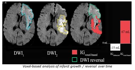 Voxel-based analysis of infarct growth / reversal over time
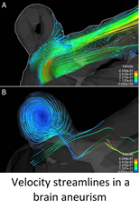 Velocity streamlines in a brain aneurism
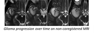 Glioma progression over time on non-coregistered MRI
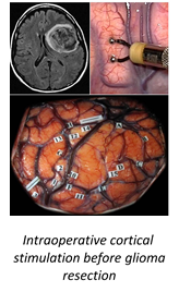 Intraoperative cortical stimulation before glioma resection
 Preoperative MRI.jpg
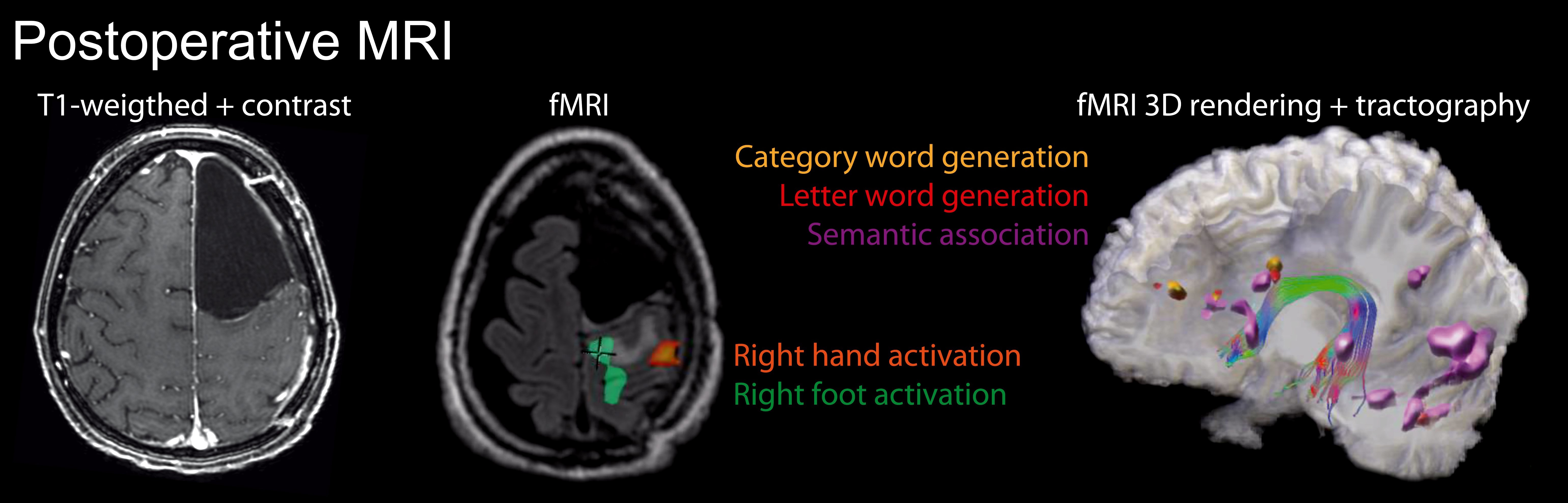 Postoperative MRI.jpg
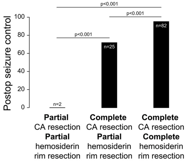 Postop seizure control
 Image Analysis Methodology
 Imaging Progression Over Time
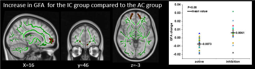 Increase of GFA
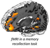 fMRI in a memory recollection task


![]()


